Graphics Gallery
Gale Rhodes
Chemistry Department
University of Southern Maine
Revised 2006/08/02
Learn how to use Swiss-PdbViewer: Work through sections 1-4 of
the Swiss-PdbViewer
Tutorial.

Topic: Nucleotides and Nucleic Acids
Several important cofactors and prosthetic groups are, or contain,
nucleotides. Nucleotides are the building blocks of nucleic acids.
Use this page to take a closer look at their structures.
Examples
I. Nucleotide Coenzymes
Following are brief descriptions of some important nucleotides or
nucleotide-containing molecules, along with links to structure files
for viewing with Swiss-PdbViewer or your favorite viewer. As you view
each structure, try to identify all of the components and bonding
groups mentioned in the description.
NOTE: Most of these structure files are derived from
crystallographic structures of proteins carrying these molecules. In
most cases, crystallographic images are not sharp enough to reveal
hydrogen atoms. Therefore, hydrogens are missing from most of these
files. A biochemist's knowledge of organic chemistry tells her or him
where the hydrogens are located. You will quickly become accustomed
to seeing biological structures without the hydrogens.
(The title of this section is Nucleotide Coenzymes. Which of the following
are better described as coenzymes, cofactors, cosubstrates, or prosthetic groups?)
- ATP, or adenosine triphosphate,
a nucleotide triphosphate composed of adenine joined by its N-9 to
C-1' of ribose by a beta linkage, with a triphosphate group on
C-5' of the ribose. As you will learn later, the triphosphate
group of ATP takes part in phosphate-group-transfer reactions.
From PDB file 1KAX.
- NAD, or nicotinamide adenine
dinucleotide, is composed of nucleosides of nicotinamide and
adenine joined at their C-5' positions by a diphosphate group. The
nicotinamide portion of NAD takes part in oxidation and reduction
reactions. From PDB file 1UDB.
- FAD, or flavin adenine
dinucleotide, is composed of adenosine and a nucleoside containing
riboflavin and the sugar alcohol ribotol. As in NAD, the two
nucleosides are joined by a diphosphate link between their 5'
carbons. Riboflavin, like nicotinamide, takes part in oxidation
and reduction reactions. From PDB file 1BV4.
- Many cofactors (including the previous three) contain AMP, but
not always with other nucleosides. An example is CoA,
coenzyme A, in which pantothenic acid and beta-mercaptoethylamine
are joined sequentially to the second phosphate group of ADP. The
sulfhydryl group of coenzyme A often carries acyl groups, joined
by a thioester link, during a variety of metabolic processes. In
this example, CoA is joined by a thioester link to the pamitolyl
group, CH3(CH2)14CO, shown in
green. You can select and display CoA or the pamitoyl group
separately. This molecule is shown as found in acyl-CoA-binding
protein from E. coli (PDB file 1ACA). The structure
was determined by NMR spectroscopy, so hydrogens are shown.
II. Nucleic Acids
Three Conformations of DNA
Following are links to structure files of the three most common
DNA conformations. After downloading each one, calculate H-bonds to
see base-pair bonding between the chains. Find AT and GC pairs from
the hydrogen-bonding patterns. Find the major and minor grooves in
each conformation. Which models are right-handed helices and which
are left-handed helices?
- B-DNA Probably the most common
DNA conformation in cells.
- A-DNA A dehydrated form of
DNA, probably not very common in cells. However, double-stranded
RNA and DNA/RNA duplexes are sometimes found
in this conformation.
- Z-DNA An unusual DNA conformation
found in the crystalline structure of certain synthetic DNAs. It
is not clear whether Z-DNA has any biological function..
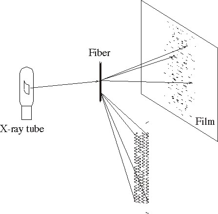
How Do We Know DNA Structure?
DNA structure was first revealed by analysis of x-ray diffraction
by DNA fibers.
- Slide 1: Fiber diffraction
experiment.
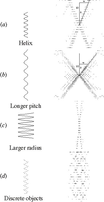
- Slide 2: Relationship
between molecular dimensions and diffraction pattern.
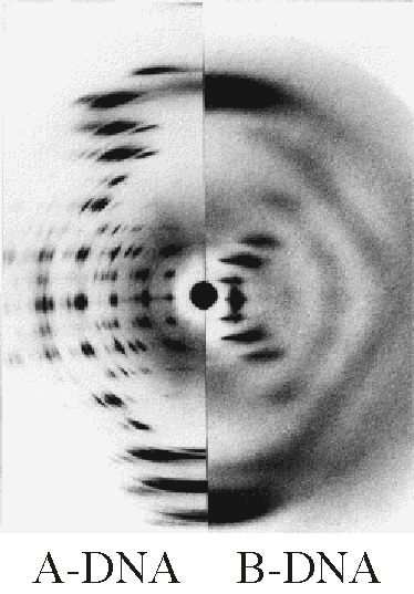
- Slide 3: Diffraction by
fibers of A- and B-DNA.
Example of Intercalation Compound
- DNA-Daunamycin Complex: This
model is an example of how molecules called intercalators
can bind to DNA. Intercalators slip in between the bases and
stretch the DNA slightly. This distortion of the shape of the
double helix can block replication or result in mistakes in
replication (mutations).
III. DNA-Protein Complexes (For Advanced Deep View Users)
Work through sections 1-6, 10, and 11 of the tutorial
before doing this exercise. Also, because water molecules are often
important participants in protein-DNA interactions, configure Deep
View to load and display water, as follows:
- Start Deep View and click Cancel on the initial
file-open dialog.
- Prefs: Loading Proteins
- Check Show Solvent (if loaded), and uncheck
Ignore solvent (WAT SOL HOH).
DNA-Eco RI Complex
Download this Deep View
project file containing a model of the restriction enzyme Eco RI
complexed to its target DNA. Superimposed on the DNA is a model of
B-DNA. Comparison of the two DNA models reveals conformational
changes forced on the DNA when Eco RI binds. Study these models as
you read about this protein-DNA complex in your text or web sources. Use blinking
(hold down control,
then press
tab) to turn the models on and off in succession.
Studying Other Protein/Nucleic-Acid Complexes
You can use the B-DNA model from Section I above to make similar
displays for other DNA-binding proteins.
- Open a DNA-protein model from the PDB.
- Open the B-DNA model from Section I.
- Display only the B-DNA and the DNA from the DNA-protein model.
Display only the backbones.
- Using the Layer Infos window to control movement,
superimpose the backbone of the B-DNA model onto the backbone of
the other DNA.
- Blink between the models to reveal the extent of
conformational change that the protein forces upon the DNA.
- Add the protein to reveal specific interactions in protein-DNA
binding.
- Use the model to help you understand more about the discussion
of this complex in your text.
- If the bound DNA looks a lot like A-DNA (Section I), try the
same operations with the A-DNA model.
If you make any such displays, please share them with me by
attaching them to email.
Download this Deep View
Project file containing seven DNA-binding proteins, each in
complex with its cognate or target DNA. In all models, the DNA is
roughly superimposed on layer #1, a dodecamer of B-DNA. The layers in
this model are listed below, with links to the PDB entry for each
model, to allow you to learn more about the models -- recall that
Deep View project files do not contain the header information for
each PDB file included. These models sample a wide range of variety
of DNA-binding protein types. Read about each one in your
biochemistry text as you explore it.
- B-DNA dodecamer (PDB 2BNA)
- 434 phage repressor, type: helix-turn-helix (PDB 2OR1)
- E. coli trp repressor, type: helix-turn-helix, indirect
readout (PDB 1TRO)
- E. coli met repressor with co-repressor, type: two-strand
antiparallel beta sheet (PDB 1CMA)
- Mouse Zif268 gene regulator protein, type: mononuclear zinc
finger (PDB 1ZAA)
- Yeast GAL4 transcription activator, type: binuclear zinc
finger (PDB
1D66)
- Mouse Max transcription factor, type helix-loop-helix with
leucine zipper (PDB 1AN2)
- Yeast GCN4 transcription activator, type: leucine zipper (PDB
1YSA)
Here are some questions you can answer for each model by exploring
with Deep View:
- Find the structural element that characterizes this particular
type of DNA-binding protein.
- Find sequence-specific interactions between protein and DNA
bases.
- Find non-specific interactions between protein and sugars or
phosphates.
- Find interactions that are mediated by water molecules.
- Does the protein interact with DNA in the major groove or the
minor groove?
- Characterize specific interactions as ionic, H-bond, polar, or
nonpolar.
- Does the protein distort the DNA double helix?
- Is the recognition site on DNA palindromic? If so, does
protein symmetry reflect sequence symmetry?
Topics List
Biochemistry
Resources