Molecular Graphics ManifestoHow should authors of texts support graphics education? |
You cannot expect authors of biochemistry textbooks to make a commitment to a specific graphics program or a specific approach to incorporation of graphics into the beginning course. But without question, the simplest and the greatest aid that authors could provide in support of graphics would be always to include the PDB file code for the models from which they derive their textbook illustrations.
For example, imagine a graphics-sophisticated student studying that typical, daunting figure that compares the specificity-determining sites of chymotrypsin, trypsin, and elastase. It is natural for this student to want to make this comparison with graphics. A PDB SearchLite search on chymotrypsin (as of 2001/01/03) produces a bewildering 300-plus hits, including models of chymotrypsin mutant forms, forms at various temperatures and other conditions, forms from a variety of organisms, and completely different proteins whose PDB file headers happen to include the word chymotrypsin.
How can students find the same model from which the figure was derived? It's very difficult even for experienced graphics and PDB users, but it's a snap if the authors routinely include PDB file codes in the captions of their illustrations. This simple practice makes it easy and natural for students to put their new graphics skills to good use.
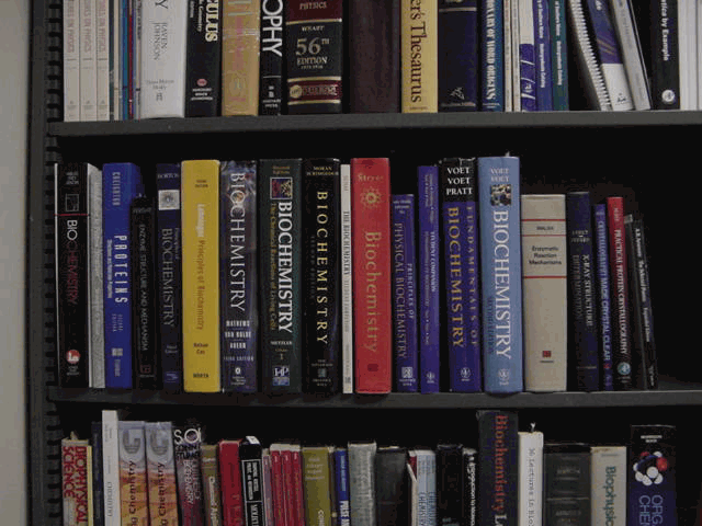
Figure: Some widely used biochemistry texts. Only one of these texts gives PDB file codes in its figure captions. Click to enlarge image.
