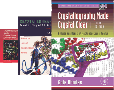
See Author's Book Sale, Below
|
Looking for a web site mentioned in CMCC-3? Web sites and servers mentioned in the book are listed under the chapter of their first mention. Chapter 1. Model and Molecule Chapter 2. An Overview of Protein
Crystallography Chapter 3. Protein
Crystals Chapter 4.
Collecting Diffraction Data Chapter 5. From Diffraction Data to Electron
Density Chapter 6. Obtaining Phases Chapter 7. Obtaining
and Judging the Molecular Model Chapter 8. A User's Guide to Crystallographic
Models Chapter
9. Other Diffraction Methods Chapter
10. Other Kinds of Macromolecular Models Chapter 11. Tools for Studying Macromolecules |
Want to learn more about structure determination? Web sites listed here are good places to expand your understanding beyond the level of CMCC-3.
|
Why Does CMCC-II Begin With A Poem?
Thanks to A. R. Ammons (1926-2001) and his publishers for allowing me to grace my book with his poem, "Phase". This poem contains images that remind me of things crystallographic, and it also includes some common terms, such as phase, that have various meanings in the English language, but quite specific meaning in crystallography. I used Ammons's images as analogies in several places in the book, and I drew contrasts between common and crystallographic meanings of terms. Can you find my allusions to the poem?
If you enjoy this poem, you
might want to learn more,
or even more, about
A.R. Ammons.
Judging Model Quality
CMCC did not have a glossary, but it does now -- one with a special purpose. This illustrated glossary confines itself to terms that are crucial to judging the quality of macromolecular models from crystallography, NMR, and homology modeling. If you don't know or can't remember the meaning of temperature factor, restraint, or threading energy, then this is the page for you.
More on Crystal Model Quality
Model Validation, Tutorial by Gerhard Kleywegt, Uppsala University
An extensive and informative tutorial that will turn you into an astute judge of model quality. Be the first kid on your block to really know whether you are working with a good model.
MolProbity: Powerful Browser-Based Model Validation Tools
Another great site for learning about -- and doing -- model validation. This site provides all the tools you need in the form of Java applets, including the latest versions of Kinemage software, either the stand-alone program Mage, or the applet KiNG. The tools at this site can grab PDB files, electron-density maps, and other resources as you work. Includes step-by-step guidance for beginners.
Real Ramachandran Plots, by Sven Hovmoeller, Stockholm University
As a tool for use in predicting protein folding, Professor Hovmoeller has compiled Ramachandran diagrams showing the actual range of phi and psi values for each of the common amino acids, as they occur in all proteins in the Protein Data Bank. These diagrams are informative, useful, and pretty as well. At Professor Hovmoeller's home page, click on Projects, and then on Protein Folding Prediction and Ramachandran Plots.
RNABase, the RNA Structure Database
Special tools for judging RNA model quality, as part of a database of all RNA structures. Links to PDB entries of RNA structures and protein-RNA complexes. Judging model quality for nucleic acids is a whole new ballgame; get your first batting and fielding practice here.Computer Graphics Tutorials for Swiss-PdbViewer and RasMol
The following tutorials are designed for beginners in macromolecular modeling. They are especially appropriate for beginning biochemistry students at the time they are learning about protein structure (after the first 5 or 6 weeks of the first semester course). The tutorials teach both the use of a computer program and basic principles of protein structure. They also introduce the Protein Data Bank. Both tutorials have been used and tested by many teachers, students, and researchers.
Tutorial for Swiss-PdbViewer (Deep View)
Learn how to use Swiss-PdbViewer by working through a self-guiding introductory tutorial. Start with simple viewing, but if you wish, go on to sophisticated comparison and analysis of structures, judging the quality of models, and homology modeling. No previous experience with molecular graphics required. You'll be a skillful modeler by the time you finish this tutorial. For version 3.1.
Swiss-PdbViewer Home
Get the free program Swiss-PdbViewer, a powerful tool for macromolecular viewing, structural analysis, comparison of models, and model building.
Tutorial for RasMol
Learn how to use this popular macromolecular viewing tool by working through a self-guiding introductory tutorial. No previous experience with molecular graphics required. For version 2.6b2.
RasMol Home
Get the free macromolecular viewer RasMol, as well as many resources for teaching structural biology using RasMol and its plug-in version Chime.
Stereo Viewing
Learn how to see the 3-D in stereo pairs while using computer graphics, reading journals, and even on projection screens during classes or seminars -- without special viewers. Good eye-muscle exercise as well.
Data Banks of Macromolecular Models
Protein Data Bank
Get models to view on your computer. The Protein Data Bank (PDB) is the world's most complete source of macromolecular models obtained by the experimental methods of diffraction and NMR. Models are provided as files of atomic coordinates in just the format you need for viewing with Swiss-PdbViewer or other graphics programs. In addition, PDB files are linked to MedLine abstracts of the papers that describe the structures.
SWISS-MODEL Repository
Currently a repository for macromolecular models obtained by automated homology modeling. This or a similar site will eventually become the PDB-like repository for all theoretical (non-crystallographic / non-NMR) models.
Data Bank of Electron-Density Maps
Enter a PDB file code and download an electron-density map. You can view maps with Swiss-PdbViewer and other graphics programs to see the quality of the map on which the structure is based. If there is no map available for your PDB file, there are two possible reasons: 1) the crystallgraphers did not deposit the x-ray data (structure factors) at the Protein Data Bank, or 2) the file is too new -- just wait awhile.
Crystallography 101
Good next step after CMCC for learning more about macromolecular crystallography. This complete short course in x-ray crystallography features excellent illustrations, interactive images, and problem solving.
A Practical Guide to Protein Crystallization
How to grow and screen crystals for X-ray Crystallography. Be sure to look at the bottom of the page for links to beautiful pictures of protein crystals.
BioSync A Resource for Synchrotron Radiation Users
This web site, maintained by the San Diego Supercomputer Center, is intended to provide technical, administrative, and logistical information to prospective synchrotron users in the field of macromolecular crystallography. The site is being developed on behalf of the Structural Biology Synchrotron Users Organization(BioSync). BioSync was formed in 1990 to promote access to synchrotron radiation for scientists whose primary research is in the field of structural biology.
Nuts and Bolts of Synchrotron X-ray Sources
Home pages for the worlds sources of high-energy X-rays for crystallography. Most sites include a virtual tour.
Kevin Cowtan's Book of Fourier
Highly informative and revealing graphical illustrations of the Fourier transform and its application to structure determination by diffraction methods. Thanks to Kevin Cowtan for allowing me to use some of his excellent images in CMCC. Many additional vivid illustrations at this site.
The Basics of NMR
Thorough introduction to NMR theory. Brief treatment of two-dimensional NMR near the end.
2D NMR Spectroscopy
Introduction to the principles of two-dimensional NMR. Emphasis on small molecules, but you need to understand these principles before pursuing a deeper understanding of multidimensional, macromolecular NMR.
Practical Applications of NMR Spectroscopy in Biochemistry
Big molecules at last! Syllabus for Professor Gordon Rule's course, Structural Biophysics 03-871, at Carnegie Mellon University. Nicely illustrated lecture outlines for most of the course topics.
BioMagResBank
A repository for data from NMR spectroscopy on proteins, peptides, and nucleic acids.
Principles of Protein Structure, Comparative Protein Modeling, and Visualisation
by Nicolas Guex and Manuel C. Peitsch
Excellent illustrated introduction to the methods and limitations of homology modeling, starting with the basics of protein structure.
ExPASy (Expert Protein Analysis System)
Powerful suite of computer tools and data bases for molecular biologists. ExPASy is the molecular biology WWW server of the Swiss Institute of Bioinformatics (SIB). This server is dedicated to the analysis of protein sequences and structures as well as identification of proteins by 2-dimensional polyacrylamide gel electrophoresis. Swiss-PdbViewer is especially designed to work with ExPASy structure tools.
Please Suggest Other Links!
If you find a web resource that you think I should list here, or if you want your own site linked to this page, please send me the URL and I will take a look. Remember that the modest goal of this page is to help CMCC readers, as well as others of similar experience, to take their next steps in learning more about macromolecular structure, structure determination, modeling, and wise use of models. See bottom of page for my email address.
Correcting Errors in CMCC-II
No book is perfect. If you find an error of any kind in CMCC-II, please notify me. I want to know about typographical errors, errors in figures, conceptual errors, even misleading or unclear passages. Your help can eliminate minor errors in future printings.
At this site, I will provide corrections for all errors, including repaired figures that you can download and paste into your copy. For error notices received to date, click the heading of this section.
Correcting Errors in CMCC-III
Appearing soon.
How Do You Use CMCC?
I will appreciate feedback about how you have used CMCC, either edition, in your research, teaching, or personal learning. Such feedback my help me to make the book and this page more useful to more readers.
Model and portion of electron-density map of bovine Rieske iron-sulfur protein (Protein Data Bank code 1rie). The electron-density map is contoured only around selected residues and the iron-sulfur center. For discussion, see Figure 11.9 and Chapter 11 of CMCC-III. Image prepared using Swiss-PdbViewer and POV-Ray. This stereo pair is designed for convergent (cross-eye) viewing. |
About Protein Crystallography
"I was captured for life by chemistry and by crystals."
Dorothy Hodgkin
Just as we see objects around us by interpreting the light reflected from them, x-ray crystallographers "see" molecules by interpreting x-rays diffracted from them. Molecules are too small to diffract visible light, but x-rays are of shorter wavelength, and thus can illuminate molecules. Each molecule diffracts too weakly to be seen, but when many molecules are aligned in a crystal, all diffracting in unison, diffracted rays are strong enough to detect and interpret. That's why protein crystals are essential to learning protein structure.
The actual molecular image obtained from crystallography is called an electron-density map (represented as a red net above, and also in this more detailed image). The crystallographer then interprets the image by building a model (balls and sticks in the figure above), fitting the known chemical contents of the protein into the map. This entails displaying map and model on a computer and fitting the model to the map. Often, the crystallographer represents frequently occuring structural elements of proteins as cartoons, like the blue and red ribbons, which are shorthands for beta sheets (blue) and alpha helices (red) of proteins. So as you look from top to bottom, the figure becomes more interpretive: first the raw image (net), then the atomic interpretation of the image (balls and sticks), and finally, cartoons (ribbons) that are shorthand for common structural elements of proteins.
Peptide page-divider image prepared with DeepView (aka Swiss-PdbViewer).
Gale Rhodes
Contact Information
Visitors Since February 2006 (release of CMCC-III)
Visitors Since January 2000 (release of CMCC-II)
